
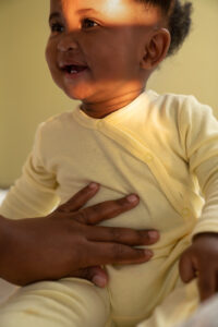
Understanding Screen Time for Children
What Does Screen Time Mean for Children?
Screen time refers to the amount of time a person spends in front of a screen, whether it’s a television, smartphone, tablet, or computer. For children, screen time includes watching videos, playing games, using educational apps, and engaging in social media.
While some screen time can be educational and beneficial, too much screen time can be harmful to a child’s overall development.
The Negative Effects of Screen Time on Children
Excessive screen time can lead to several negative consequences for your child, affecting their physical, mental, and social well-being.
Here are some key areas where screen time can have a detrimental impact:
1. Physical Health Issues
Spending too much time in front of screens can lead to a sedentary lifestyle, contributing to obesity and other health problems. Children who spend more time sitting and engaging with screens do not want to participate in physical activities, which are essential for healthy growth and development.
2. Sleep Disruptions
Excessive screen time on mobile phones, tablets, laptops, and other devices, especially before bedtime, can interfere with a child’s sleep. The blue light emitted from screens can disrupt the production of melatonin, a hormone responsible for sleep regulation. This can lead to difficulty falling asleep, poor sleep quality, and daytime fatigue.
3. Impact on Vision
Prolonged exposure to screens can strain a child’s eyes, leading to issues like dry eyes, headaches, and even myopia (nearsightedness). Regular breaks and limiting screen time are very important to protect your child’s vision.
4. Delayed Language Development
Young children need face-to-face interactions to develop their language skills. Excessive screen time can limit these interactions, leading to delayed speech and language development in toddlers and young children.
5. Attention and Learning Issues
Children who spend too much time on screens may struggle with attention and concentration. This can affect their ability to focus in school and complete tasks, potentially leading to academic challenges.
6. Social and Emotional Development
Screen time can impact a child’s social skills and emotional development. Children who spend more time on screens may have less time for social interactions, leading to difficulties in maintaining and creating relationships.
குழந்தை வளர்ச்சியில் திரை நேரத்தின் தாக்கம்: பெற்றோருக்கான வழிகாட்டுதல்
இன்றைய டிஜிட்டல் சகாப்தத்தில், குழந்தைகள் முன்
எப்போதும் இல்லாத அளவுக்கு மனிதர்களுடன் அதிக நேரம் செலவிடுகிறார்கள்.
தொலைக்காட்சிகள், ஸ்மார்ட்போன்கள், டேப்லெட்டுகள் மற்றும் கணினிகள் முதல், திரைகள்
அனைவரின் அன்றாட வாழ்விலும் குறிப்பிடத்தக்க பகுதியாக மாறிவிட்டன. ஆம்,
தொழில்நுட்பம் பல நன்மைகளை வழங்குகிறது, ஆனால் அதிகப்படியான திரை நேரம் குழந்தை
வளர்ச்சியில் எதிர்மறையான விளைவுகளை ஏற்படுத்தும்.
இந்த வலைப்பதிவில், குழந்தைகளின் திரை நேரத்தின்
தாக்கத்தை ஆராய்வோம், ஆம்பூர் மற்றும் திருப்பத்தூர் மாவட்டங்களில் உள்ள
பெற்றோருக்கு வழிகாட்டுதல்களை வழங்குவோம், மேலும் உங்கள் குழந்தையின் திரை நேரத்தை
எவ்வாறு திறம்பட நிர்வகிப்பது என்பதற்கான சில பரிந்துரைகளை வழங்குவோம்.
குழந்தைகளுக்கான திரை நேரத்தைப் புரிந்துகொள்வது
குழந்தைகளுக்கு
திரை நேரம் என்றால் என்ன?
திரை நேரம் என்பது ஒரு நபர் ஒரு தொலைக்காட்சி,
ஸ்மார்ட்போன், டேப்லெட் அல்லது கணினியாக இருந்தாலும், திரையின் முன் செலவழிக்கும்
நேரத்தைக் குறிக்கிறது. குழந்தைகளுக்கான திரை நேரம் என்பது வீடியோக்களைப் பார்ப்பது,
கேம்களை விளையாடுவது, கல்வி சார்ந்த பயன்பாடுகளைப் பயன்படுத்துவது மற்றும் சமூக
ஊடகங்களில் ஈடுபடுவது ஆகியவை அடங்கும். சில திரை நேரம் கல்வி மற்றும் நன்மை
பயக்கும் அதேசமயம், அதிக திரை நேரம் குழந்தையின் ஒட்டுமொத்த வளர்ச்சிக்கு தீங்கு
விளைவிக்கும்.
குழந்தைகள் மீது திரை நேரத்தின் எதிர்மறை விளைவுகள்
அதிகப்படியான திரை நேரம் உங்கள் பிள்ளைக்கு பல
எதிர்மறையான விளைவுகளுக்கு வழிவகுக்கும், இது அவர்களின் உடல், மன மற்றும் சமூக
நலனை பாதிக்கும்.
திரை நேரம் தீங்கு விளைவிக்கும்
சில முக்கிய பகுதிகள் இங்கே:
1. உடல் நலப் பிரச்சினைகள்: திரைகளுக்கு முன்னால்
அதிக நேரம் செலவிடுவது உடல் பருமன் மற்றும் பிற உடல்நலப் பிரச்சினைகளுக்கு
பங்களிக்கும், உட்கார்ந்த வாழ்க்கை முறைக்கு வழிவகுக்கும். அதிக நேரம் உட்கார்ந்து
திரையில் ஈடுபடும் குழந்தைகள் ஆரோக்கியமான வளர்ச்சி மற்றும் வளர்ச்சிக்கு அவசியமான
உடல் செயல்பாடுகளில் பங்கேற்க விரும்புவதில்லை.
2. தூக்கக் கோளாறுகள்: மொபைல், டேப்லெட், லேப்டாப்
போன்றவற்றில் அதிக நேரம் திரையிடுவது, குறிப்பாக தூங்கும் முன், குழந்தையின்
தூக்கத்தில் குறுக்கிடலாம். திரைகளில் இருந்து வெளிப்படும் நீல ஒளியானது தூக்கத்தை
ஒழுங்குபடுத்தும் மெலடோனின் என்ற ஹார்மோனின் உற்பத்தியை சீர்குலைக்கும்.
இது தூங்குவதில் சிரமம், மோசமான தூக்கத்தின் தரம்
மற்றும் குழந்தைகள் பகல்நேர சோர்வை எதிர்கொள்ளலாம்.
3. பார்வை மீதான தாக்கம்: திரைகளில் நீண்ட நேரம்
வெளிப்படுவது குழந்தையின் கண்களைக் கஷ்டப்படுத்தலாம், இது உலர் கண்கள், தலைவலி
மற்றும் கிட்டப்பார்வை (கிட்டப்பார்வை) போன்ற பிரச்சினைகளுக்கு வழிவகுக்கும். உங்கள் குழந்தையின்
பார்வையைப் பாதுகாக்க, வழக்கமான இடைவெளிகளும், திரை நேரத்தைக் கட்டுப்படுத்துவதும்
மிகவும் முக்கியம்.
4. தாமதமான மொழி வளர்ச்சி: சிறு குழந்தைகள் தங்கள் மொழித்
திறனை வளர்த்துக் கொள்ள நேருக்கு நேர் தொடர்பு கொள்ள வேண்டும். அதிகப்படியான திரை
நேரம் இந்த தொடர்புகளை மட்டுப்படுத்தலாம், இது குழந்தைகள் மற்றும் சிறு
குழந்தைகளின் பேச்சு மற்றும் மொழி வளர்ச்சி தாமதத்திற்கு வழிவகுக்கும்.
5. கவனம் மற்றும் கற்றல் சிக்கல்கள்: திரையில் அதிக நேரம்
செலவழிக்கும் குழந்தைகள் கவனம் மற்றும் கவனத்துடன் போராடலாம். இது அவர்களின்
பள்ளியில் கவனம் செலுத்தும் திறனைப் பாதிக்கலாம் மற்றும் பணிகளை முடிக்கலாம், இது
கல்வி சார்ந்த சவால்களுக்கு வழிவகுக்கும்.
6. சமூக மற்றும் உணர்ச்சி வளர்ச்சி: திரை நேரம் குழந்தையின்
சமூக திறன்கள் மற்றும் உணர்ச்சி வளர்ச்சியை பாதிக்கலாம். திரையில் அதிக நேரம்
செலவழிக்கும் குழந்தைகளுக்கு சமூக தொடர்புகளுக்கு குறைவான நேரம் இருக்கலாம், இது
உறவுகளை பராமரிப்பதிலும் உருவாக்குவதிலும் சிரமங்களுக்கு வழிவகுக்கும்.
வயதின்
அடிப்படையில் திரை நேர பரிந்துரைகள்
குழந்தையின் ஆரோக்கியமான மற்றும் சமநிலையான திரை நேரத்தை உறுதிசெய்ய, வயதுக்கு
ஏற்ற வழிகாட்டுதல்களைப் பின்பற்றுவது முக்கியம். நோய் கட்டுப்பாடு மற்றும் தடுப்பு
மையங்கள் (CDC) https://www.cdc.gov/ மற்றும் பிற சுகாதார நிறுவனங்கள் குழந்தையின்
வயதை அடிப்படையாகக் கொண்டு திரை நேரம் குறித்த பரிந்துரைகளை வழங்குகின்றன:
– 18 மாதங்களுக்கு கீழ் உள்ள குழந்தைகள்:
குடும்ப உறுப்பினர்களுடன் வீடியோ அரட்டையைத் தவிர, திரை நேரத்தைத் தவிர்க்கவும்
– 18-24 மாத வயதுடைய குழந்தைகள்: உயர்தர கல்வி
உள்ளடக்கத்தில் கவனம் செலுத்தி, குறைந்த நேர இடைவெளியில் டிஜிட்டல் மீடியாவை
அறிமுகப்படுத்துங்கள். திரை நேரத்தில் பெற்றோர்கள் எப்போதும் தங்கள் குழந்தைகளைப்
பார்த்து அவர்களுடன் பழக வேண்டும். (பெற்றோர் தலையீடு இன்றியமையாதது)
– 2-5 வயதுடைய குழந்தைகள்: ஒரு நாளைக்கு 1
மணிநேரத்திற்கு மிகாமல் திரை நேரத்தைக் கட்டுப்படுத்துங்கள். ஊடாடும் மற்றும்
கல்வி உள்ளடக்கத்தை ஊக்குவிப்பது நல்லது.
– 6 வயது மற்றும் அதற்கு மேற்பட்ட குழந்தைகள்:
திரை நேரத்தில் எதிர்பார்க்கப்படும் வரம்புகளை அமைக்கவும், அது தூக்கம், உடல்
செயல்பாடு மற்றும் பிற ஆரோக்கியமான நடத்தைகளில் தலையிடாது என்பதை
உறுதிப்படுத்துகிறது. படைப்பாற்றல் மற்றும் கற்றலை ஊக்குவிக்கும் செயல்களில்
அவர்களை எப்போதும் ஊக்குவிக்க வேண்டும்
அதிக திரை நேரம் குழந்தைகளுக்கு மோசமாக
இருப்பதற்கான 10 காரணங்கள்
அதிக திரை நேரம் தீங்கானது என்பதற்கான குறிப்பிட்ட காரணங்களைப்
புரிந்துகொள்வது, அதை நிர்வகிப்பது குறித்து பெற்றோர்கள் சரியான முடிவுகளை எடுக்க
உதவும் .
உங்கள் குழந்தையின் வளர்ச்சிக்கு திரை நேரத்தைக் கட்டுப்படுத்துவது மிகவும்
முக்கியமானது என்பதற்கான 10 காரணங்கள் இங்கே:
1. உடல் செயல்பாடுகளை குறைக்கிறது: திரையில்
அதிக நேரம் செலவிடும் குழந்தைகள் உடல் விளையாட்டில் ஈடுபடுவது குறைவு, இது
உட்கார்ந்த வாழ்க்கை முறை மற்றும் தொடர்புடைய உடல்நலப் பிரச்சினைகளுக்கு
வழிவகுக்கும்.
2. தூக்கத்தின் தரத்தில் ஏற்படும் பாதிப்புகள்:
மேலே குறிப்பிட்டுள்ளபடி, திரைகளில் இருந்து வெளிவரும் நீல ஒளியானது தூக்க முறைகள்
மற்றும் அட்டவணைகளை சீர்குலைத்து, குழந்தைகளுக்குத் தேவையான ஓய்வு கிடைப்பதை
கடினமாக்குகிறது.
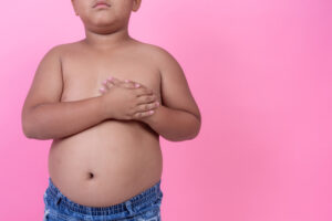
3. உடல் பருமனின் அபாயத்தை அதிகரிக்கிறது: திரை
நேரத்தில் ஆரோக்கியமற்ற உணவுப் பழக்கங்களுடன் இணைந்து செயல்படாத வாழ்க்கை முறை எடை
அதிகரிப்பு மற்றும் உடல் பருமனுக்கு வழிவகுக்கும்.
4. மன ஆரோக்கியத்தை பாதிக்கிறது: அதிக திரை
நேரம் கவலை, மனச்சோர்வு அல்லது பிற மனநலப் பிரச்சினைகள் போன்ற பல அறிவாற்றல்
சிக்கல்களுக்கு வழிவகுக்கும், குறிப்பாக சற்று வயதான குழந்தைகள் மற்றும் இளம்
பருவத்தினருக்கு.
5. கல்வித் திறனைப் பாதிக்கிறது: திரையில் அதிக
நேரம் செலவழிக்கும் குழந்தைகள் கவனத்துடனும கவனத்துடனும் போராடலாம், இது கல்விச்
சாதனையில் சிக்கல்களுக்கு வழிவகுக்கும்.
6. குறைக்கப்பட்ட சமூக தொடர்பு: அதிகப்படியான
திரை நேரம் நேருக்கு நேர் தொடர்புகளை கட்டுப்படுத்தலாம், இது குழந்தைகளுக்கு சமூக
திறன்களை வளர்ப்பதை கடினமாக்குகிறது.
7. மொழியியல் வளர்ச்சியில் தாமதங்கள்: இளம்
குழந்தைகளுக்கு மொழித் திறன்களை வளர்த்துக் கொள்ள வாய்மொழி தொடர்புகள் மற்றும்
மனிதத் தொடர்பு தேவை, இது அதிக திரை நேரத்தால் தடைபடும்.
8. நடத்தை சிக்கல்கள்: பல ஆய்வுகள் திரைகளில்
அதிக நேரம் செலவிடும் குழந்தைகள் ஆக்கிரமிப்பு மற்றும் கீழ்ப்படியாமை போன்ற நடத்தை
சார்ந்த பிரச்சனைகளை வெளிப்படுத்தும் வாய்ப்புகள் அதிகம் என்று காட்டுகின்றன.
9.
படைப்பாற்றலைத் தடுக்கிறது: ஒரு குழந்தையின் வளர்ச்சிக்கு
படைப்பாற்றல் அவசியம், இது அவர்களுக்கு வெளியே சிந்திக்க உதவுகிறது, ஆனால் அதிக
திரை நேரம் கண்டுபிடிப்பு மற்றும் கற்பனை செயல்பாடுகளுக்கான வாய்ப்புகளை
குறைக்கலாம்.
10. பொருத்தமற்ற உள்ளடக்கத்தின் வெளிப்பாட்டை
அதிகரிக்கிறது: பெரியவர்களின் சரியான மேற்பார்வை இல்லாமல், குழந்தைகள் ஆன்லைனில்
வன்முறை, பொருத்தமற்ற மற்றும் தீங்கு விளைவிக்கும் உள்ளடக்கத்திற்கு ஆளாகலாம்.
டீனேஜ் மூளையில் திரை நேரத்தின் விளைவுகள்
குழந்தைகள் வளரும்போது, திரையின் நேரத்தின் தாக்கம் குறிப்பாக பதின்ம
வயதினருக்கு இன்னும் அதிகமாகத் தெரியும். டீனேஜ் மூளை இன்னும் வளர்ந்து வருகிறது,
அதிகப்படியான திரை நேரம் இந்த செயல்முறையை பல்வேறு வழிகளில் பாதிக்கலாம்:
1.
நரம்பியல் வளர்ச்சி: அதிக நேரத்தை திரையில் செலவிடும்
பதின்வயதினர் மூளையின் அமைப்பில், குறிப்பாக கவனம், நினைவாற்றல் மற்றும் முடிவெடுப்பது
தொடர்பான பகுதிகளில் மாற்றங்களை அனுபவிக்கலாம்.
2.
ரிஸ்க்-டேக்கிங் பிஹேவியர்: முடிவெடுப்பதற்கும் உந்துவிசைக்
கட்டுப்பாட்டிற்கும் பொறுப்பான ப்ரீஃப்ரொன்டல் கோர்டெக்ஸ் (மூளையின் ஆளுமை மையம்)
இன்னும் பதின்ம வயதினரிடையே உருவாகி வருகிறது. அதிக திரை நேரம், குறிப்பாக சமூக
ஊடகங்கள் அல்லது கேமிங்கை உள்ளடக்கியது, அதிக ரிஸ்க் எடுக்கும் நடத்தைக்கு
வழிவகுக்கும்.
3.
உணர்ச்சிக் கட்டுப்பாடு: டிஜிட்டல் மீடியாவின் தொடர்ச்சியான
வெளிப்பாடு, உணர்ச்சிகளைக் கட்டுப்படுத்தும் இளைஞனின் திறனைப் பாதிக்கலாம், இது
அதிகரித்த மன அழுத்தம், பதட்டம் மற்றும் மனச்சோர்வுக்கு வழிவகுக்கும்.
4.
தூக்கமின்மை: டீனேஜர்கள் குறிப்பாக தூக்கத்தில் நீல ஒளியின்
விளைவுகளுக்கு ஆளாகிறார்கள், இது தூக்கமின்மை மற்றும் தொடர்புடைய உடல்நலப்
பிரச்சினைகளுக்கு வழிவகுக்கிறது.
5.
அடிமையாதல்: டீன் ஏஜ் மூளையில் உள்ள வெகுமதி அமைப்பு
மிகவும் உணர்திறன் கொண்டது, குறிப்பாக சமூக ஊடகங்களில் டீனேஜர்கள் திரை
அடிமைத்தனத்திற்கு மிகவும் பாதிக்கப்படுகின்றனர்.
குழந்தைகளுக்கான திரை நேர செயல்பாடுகள்
சரி, திரை நேரத்தைக் கட்டுப்படுத்துவது முக்கியம், ஆரோக்கியமான திரை நேரம்
மற்றும் வளர்ச்சியை ஊக்குவிக்கும் மாற்று செயல்பாடுகளை குழந்தைகளுக்கு வழங்குவதும்
சமமாக முக்கியமானது.
உங்கள் குழந்தை வளரவும் கற்றுக்கொள்ளவும் உதவும் சில திரை இல்லாத செயல்பாடுகள்
இங்கே உள்ளன:
1.
வெளிப்புற விளையாட்டு: உங்கள் பிள்ளை சைக்கிள் ஓட்டினாலும்,
விளையாட்டாக இருந்தாலும், பூங்காவில் ஓடினாலும், வெளியில் விளையாடுவதை
ஊக்குவிக்கவும். உடல் செயல்பாடு ஒட்டுமொத்த ஆரோக்கியத்திற்கும் நல்வாழ்விற்கும்
இன்றியமையாதது.
2.
படித்தல்: சிறு வயதிலேயே உங்கள் பிள்ளைக்கு புத்தகங்களை
அறிமுகப்படுத்தி, வாசிப்பின் மகிழ்ச்சியைக் கற்றுக்கொள்ளட்டும். புத்தகங்கள்
அவர்களை வெவ்வேறு உலகங்களுக்கு கொண்டு செல்லவும், அவர்களின் சொற்களஞ்சியத்தை
விரிவுபடுத்தவும், அவர்களின் செறிவை மேம்படுத்தவும் முடியும்.
3.
ஆக்கப்பூர்வமான கலைகள் மற்றும் கைவினைப்பொருட்கள்: கலை
மற்றும் கைவினை நடவடிக்கைகளில் உங்கள் குழந்தையை ஈடுபடுத்துங்கள். வரைதல், ஓவியம்
மற்றும் DIY கள் அவர்களின் படைப்பாற்றலை மேம்படுத்துவதோடு மோட்டார் திறன்களை
மேம்படுத்தலாம்.
4.
குடும்ப நேரம்: குடும்பமாக தரமான நேரத்தை செலவிடுங்கள்.
போர்டு கேம்களை விளையாடுங்கள், ஒன்றாக சமைக்கலாம், ஒன்றாக வீட்டு வேலைகளில்
ஈடுபடலாம் அல்லது குடும்பத்திற்கு ஏற்ற திரைப்படத்துடன் இரவு திரைப்படம்
பார்க்கலாம்.
5.
கல்வி சார்ந்த விளையாட்டுகள்: உங்கள் குழந்தை திரை நேரம்
பிடித்தால், ஊடாடும் மற்றும் கற்றலை ஊக்குவிக்கும் கல்வி விளையாட்டுகளை
அவர்களுக்கு அறிமுகப்படுத்துங்கள். (நேரங்களைக் கட்டுப்படுத்துவதை
உறுதிப்படுத்திக் கொள்ளுங்கள்.
6.
இசை மற்றும் நடனம்: உங்கள் பிள்ளையை இசைக்கருவி, பாடுதல்
அல்லது நடனம் கற்றுக் கொள்ள ஊக்குவிக்கவும். இந்த நடவடிக்கைகள் அவர்களின்
அறிவாற்றல் வளர்ச்சிக்கு வேடிக்கையாகவும் பயனுள்ளதாகவும் இருக்கும்.
வயது விளக்கப்படத்தின்படி திரை நேரப் பரிந்துரைகள்
பெற்றோர்கள் தங்கள் குழந்தையின் திரை நேரத்தை நிர்வகிப்பதை எளிதாக்க, வயது
அடிப்படையில் பரிந்துரைக்கப்பட்ட எளிய விளக்கப்படம் இதோ:
|
வயது குழு |
ஒரு நாளைக்கு பரிந்துரைக்கப்படும் திரை நேரம் |
பெற்றோருக்கான உதவிக்குறிப்புகள் |
|
18 மாதங்களுக்கு கீழ் |
வீடியோ அரட்டையைத் தவிர, திரை நேரத்தைத் தவிர்க்கவும் |
நிஜ உலக தொடர்புகளில் கவனம் செலுத்துங்கள் |
|
18-24 மாதங்கள் |
உயர்தர உள்ளடக்கத்துடன் வரையறுக்கப்பட்ட திரை நேரம் |
திரை நேரத்தில் உங்கள் குழந்தையைப் பார்த்து அவருடன் தொடர்பு கொள்ளவும் |
|
2-5 ஆண்டுகள் |
ஒரு நாளைக்கு 1 மணிநேரத்திற்கு மேல் திரை நேரம் இல்லை |
கல்வி உள்ளடக்கம் மற்றும் செயலில் உள்ள நேரம் ஆகியவற்றை முதன்மைப்படுத்தவும் |
|
6-12 ஆண்டுகள் |
நிலையான வரம்புகள், உடல் செயல்பாடுகளுக்கு முன்னுரிமை கொடுங்கள் |
பொழுதுபோக்கு மற்றும் வெளிப்புற விளையாட்டுகளை ஊக்குவிக்கவும் |
|
13-18 ஆண்டுகள் |
திரை நேரத்தைக் கண்காணித்து வரம்பிடவும், குறிப்பாக இரவில் |
அதிகப்படியான திரை நேரத்தின் |
உங்கள் குழந்தையின் ஆரோக்கியமான திரை நேரத்திற்கு நடவடிக்கை எடுப்பது.
உங்கள் பிள்ளையின் திரை நேரத்தை நிர்வகிப்பது அவர்களின் ஒட்டுமொத்த வளர்ச்சிக்கு
அவசியம். சாத்தியமான எதிர்மறையான விளைவுகளைப் புரிந்துகொள்வதன் மூலமும், வயதுக்கு
ஏற்ற வழிகாட்டுதல்களைப் பின்பற்றுவதன் மூலமும், உங்கள் குழந்தை ஆரோக்கியமாகவும்,
மகிழ்ச்சியாகவும், பல்துறை ரீதியாகவும் வளர்வதை உறுதிப்படுத்த உதவலாம்.
உங்கள் பிள்ளையின் திரை நேரத்தை நிர்வகிப்பது அவர்களின் ஒட்டுமொத்த
வளர்ச்சிக்கு அவசியம். சாத்தியமான எதிர்மறையான விளைவுகளைப் புரிந்துகொள்வதன்
மூலமும், வயதுக்கு ஏற்ற வழிகாட்டுதல்களைப் பின்பற்றுவதன் மூலமும், உங்கள் குழந்தை
ஆரோக்கியமாகவும், மகிழ்ச்சியாகவும், பல்துறை ரீதியாகவும் வளர்வதை உறுதிப்படுத்த
உதவலாம்.
நினைவில் கொள்ளுங்கள், முக்கியமானது சமநிலை; உடல் செயல்பாடு, சமூக தொடர்புகள்
மற்றும் ஆக்கப்பூர்வமான விளையாட்டு ஆகியவற்றை ஊக்குவிக்கும் அதே வேளையில்
தொழில்நுட்பத்தின் பலன்களை உங்கள் குழந்தை அனுபவிக்க அனுமதிக்கிறது.
ஆம்பூர் NU மருத்துவமனைகளில், இன்றைய டிஜிட்டல் உலகில் பெற்றோர்கள்
எதிர்கொள்ளும் சவால்களை நாங்கள் புரிந்துகொள்கிறோம். உங்கள் குழந்தையின்
வளர்ச்சியைப் பற்றி உங்களுக்குக் கவலைகள் இருந்தாலோ அல்லது திரை நேரத்தை
நிர்வகிப்பதற்கான வழிகாட்டுதல் தேவைப்பட்டால், நாங்கள் உதவ இருக்கிறோம்.
டாக்டர் பழனி ராஜன் பி, சீனியர் ஆலோசகர் குழந்தை நல மருத்துவர் &
நியோனாட்டாலஜிஸ்ட், குழந்தைகள் நலப் பிரிவு, KM NU மருத்துவமனை, ஆம்பூர்,
உங்களுக்கு ஏதேனும் கேள்விகள் அல்லது கவலைகள் இருந்தால், ஆலோசனைகள் கிடைக்கும்.
உங்கள் குழந்தையின் ஆரோக்கியம் மற்றும் நல்வாழ்வை உறுதிப்படுத்த ஆம்பூரில்
உள்ள எங்கள் மருத்துவமனைக்குச் செல்லவும்.
உங்கள் குழந்தையின் திரை நேரம் மற்றும் ஒட்டுமொத்த வளர்ச்சியைப் பற்றி
விவாதிக்க, ஆம்பூரில் உள்ள KM NU மருத்துவமனைகளில் டாக்டர் பழனிராஜனுடன் இன்று
சந்திப்பைத் திட்டமிடலாம்.
ஒன்றாக, உங்கள் குழந்தை ஒரு சீரான மற்றும் ஆரோக்கியமான சூழலில் செழிக்க
உதவலாம்.
Appendix Removal Surgery: Understanding Its Causes & Symptoms
Appendicectomy aka Appendectomy is the surgical removal of the appendix because of the medical condition Appendicitis. When your appendix is infected or is injured, your body will produce a response (i.e. inflammation).
While it can be a sudden and painful experience, understanding the causes, symptoms, and treatment can help you recognize the condition and seek timely medical care.
Causes of Appendicitis
Appendicitis is primarily caused by a blockage in the appendix, a small, finger-shaped pouch that extends from the large intestine. This blockage can be caused by:
- Fecal matter: Hardened stool can obstruct the appendix’s opening.
- Foreign objects: Rarely, small objects can become lodged in the appendix.
- Infection: Inflammation in the digestive system can lead to swelling of the appendix.
- Tumors: Less commonly, growths within the appendix can cause a blockage.
When the appendix gets blocked, bacteria can build up and cause inflammation, leading to appendicitis.
Symptoms of Appendicitis
Recognizing the symptoms of appendicitis is necessary for timely treatment. While symptoms can vary, common indicators include:
- Belly pain: This is often the first and most noticeable symptom. The pain usually starts around the navel and then moves to the lower right abdomen.
- Loss of appetite: As the inflammation worsens, you may lose interest in food.
- Nausea and vomiting: These symptoms often accompany abdominal pain.
- Low-grade fever: A mild fever may develop as the infection progresses.
- Tenderness in the lower right abdomen: The area may be sensitive to touch.
- Prevention of rupture: If left untreated, the appendix can rupture, leading to a potentially life-threatening infection called peritonitis.
- Relief from pain: Removing the inflamed appendix eliminates the source of pain and discomfort.
- Improved overall health: By preventing complications, appendectomy contributes to overall well-being.
- Open appendectomy: This involves a larger incision in the lower right abdomen to access and remove the appendix.
- Laparoscopic appendectomy: This minimally invasive surgical procedure uses small cuts (incisions) and a laparoscope (a thin, lighted tube with a camera) to visualize and remove the appendix.
- Pain at the incision site
- Nausea and vomiting
- Fatigue
- Constipation or diarrhoea
- These symptoms usually subside within a few days. However, if you experience severe or persistent pain, fever, or other concerning symptoms, it’s important to contact your doctor.
It’s important to note that these symptoms can also be associated with other conditions, so it’s essential to consult a healthcare professional for a proper diagnosis.
Benefits of Appendectomy
While the appendix itself is not considered essential for survival, removing it becomes necessary to prevent serious complications. The benefits of appendectomy include:
·
How Common is Appendix Removal?
Appendectomy is one of the most common emergency surgeries performed. While the exact statistics vary, it’s estimated that around 9% to 10% of the population will experience appendicitis at some point in their lives. The most common age range for appendicitis falls under 10 to 20 years.
Appendix Removal Reasons
The primary reason for an appendectomy is appendicitis. This occurs when the appendix becomes inflamed and infected, often due to a blockage. Other less common reasons for removing the appendix might include cancer or diverticulitis. However, in most cases, the appendix is removed due to appendicitis.
Appendix Removal Surgery Procedure
There are two main types of appendectomy:
The choice between these methods depends on factors such as the severity of appendicitis, the surgeon’s expertise, and the patient’s overall health.
It is a relatively safe operation. Additionally, Laparoscopic appendectomy is the preferred treatment that is also safe for pregnant women, individuals with obesity, children, and older patients (senior citizens).
How Long Does Appendix Removal Surgery Take?
The duration of an appendectomy varies depending on several factors, including the type of surgery performed and any complications encountered.
Generally, a laparoscopic appendectomy tends to be shorter than an open surgical procedure.
On average, the surgery can last up to an hour. However, preparation and recovery time should also be considered.
Recovery Time After Appendix Removal
Recovery time after an appendectomy depends on the type of surgery performed and the individual’s overall health.
Laparoscopic surgery typically allows for a quicker recovery compared to open surgery.
Most patients can resume normal activities within a few weeks, but following your doctor’s recommendations is essential.
After Surgery Effects
Common after-effects of an appendectomy include:
Can Appendix Removal Cause Bowel Problems?
While rare, it’s possible to experience bowel problems after an appendectomy. These issues can include constipation, diarrhea, or changes in bowel habits. Most of the time, these problems are temporary and resolve on their own. However, if you have persistent or severe bowel issues, consult your doctor.
In most cases, appendectomy is a safe and effective procedure with minimal long-term health consequences. However, as with any surgery, there is a risk of complications, such as infection, bleeding, or injury to nearby organs. These complications are relatively uncommon but should be discussed with your doctor before the procedure.
Can Appendix Removal Surgery Affect Fertility?
The appendix is not involved in reproductive function. Also, there is no scientific evidence to suggest that appendectomy causes infertility in either men or women.
Conclusion:
If you experience symptoms of appendicitis, such as abdominal pain, nausea, vomiting, or fever, it is time you seek medical attention promptly. Early diagnosis and treatment can prevent complications.
KM NU Hospitals is a multispecialty hospital situated in Ambur. Our hospital has many facilities to carry out an appendectomy and Laparoscopic Appendectomy. To avail the best appendix removal surgical treatment in Ambur, visit our experts at KM NU Hospitals today!
References:
NCBI: https://www.ncbi.nlm.nih.gov/books/nbk580514/
NHS UK: https://www.nhs.uk/conditions/appendicitis/treatment/
Chest Congestion and Fever in Children: Understanding its Causes, Symptoms, and Treatment
Chest congestion and fever are common concerns for parents, especially in young children. These symptoms often signal a respiratory infection, ranging from mild colds to more severe conditions like pneumonia.
This blog post aims to shed light on various aspects of chest congestion and infections in children, including symptoms, causes, and treatment options, to help parents recognize and manage these issues effectively.
Chest Congestion and Fever in Children
Chest congestion in children is typically a result of mucus build-up in the airways, which can make breathing difficult and lead to coughing. When accompanied by fever, it often indicates that the body is fighting off an infection. Fever is the body’s natural response to infection and is common in children with chest congestion.
- Common Causes:
The most frequent causes of chest congestion and fever in children include viral infections like the common cold, influenza, and respiratory syncytial virus (RSV). Bacterial infections, such as bronchitis or pneumonia, can also cause these symptoms.
- When to Worry:
While mild chest congestion and low-grade fever may not be cause for immediate concern, persistent or severe symptoms, high fever, difficulty breathing, or lethargy warrant a visit to the doctor.
Coughing is one of the primary symptoms of a chest infection in children. Chest infections can cause significant discomfort and may disrupt sleep and daily activities.
-
Viral vs. Bacterial: Chest infections in kids are commonly viral, which means antibiotics are not effective. However, bacterial infections like pneumonia require antibiotic treatment.
-
Duration: A cough associated with a chest infection can last for several weeks, even after the infection has cleared. It’s important to monitor the child for any signs of worsening symptoms, such as increased difficulty breathing or high fever.
Are Chest Infections in Babies Contagious?
Yes, chest infections in babies can be contagious, especially if they are caused by a viral infection like RSV, which is highly contagious among infants and young children.
-
Transmission: These infections spread through respiratory droplets when an infected person coughs or sneezes. Close contact, such as kissing or touching a baby’s face after handling a contaminated object, can also spread the infection.
-
Prevention: To prevent the spread of chest infections, practice good hygiene, such as frequent handwashing, keeping sick children home from daycare, and avoiding close contact with infected individuals.
Are Chest Infections in Toddlers Common?
Chest infections in toddlers are quite common, particularly during the colder months when viral infections are more prevalent.
Toddlers are at a higher risk of acquiring infection due to their developing immune systems and frequent exposure to other children in daycare or preschool settings who may have already been infected.
The most common types of chest infections in toddlers include bronchiolitis, caused by RSV, and pneumonia.
Symptoms and Signs of Chest Infection in Kids:
Recognizing the symptoms of a chest infection in kids is crucial for timely treatment. The symptoms can vary depending on the type and severity of the infection.
-
Cough: Persistent coughing is a hallmark symptom, often accompanied by wheezing or a rattling sound in the chest.
-
Fever: A high fever may indicate a bacterial infection like pneumonia.
-
Breathing Difficulties: Rapid or labored breathing, flaring nostrils, or chest retractions (where the skin pulls in around the ribs) are signs of severe infection and require immediate medical attention.
-
Lethargy: A significant decrease in activity levels or a lack of interest in eating or playing can be a sign of a more serious infection.
Symptoms in Toddlers and Babies:
The symptoms of chest infections in toddlers and babies can be subtle and may differ slightly from those in older children.
-
Babies: Symptoms in babies may include irritability, poor feeding, nasal flaring, and grunting sounds when breathing. Babies with severe chest infections may also have a bluish tint to their lips or face, indicating a lack of oxygen.
-
Toddlers: Toddlers may show symptoms like persistent coughing, wheezing, difficulty breathing, and high fever. They might also be unusually tired or clingy.
Symptoms of Infection in Children
Infections in children can present with a variety of symptoms, depending on the type and location of the infection.
-
General Symptoms: Fever, lethargy, irritability, and loss of appetite are common signs of infection in a child.
-
Respiratory Infections: Symptoms include cough, congestion, runny nose, and difficulty breathing.
-
Gastrointestinal Infections: These may cause vomiting, diarrhoea, and abdominal pain.
-
Ear Infections: Look for symptoms such as ear pain, tugging at the ear, and difficulty hearing.
Symptoms of Respiratory Infections in Children to Watch For:
Respiratory infections in children can range from mild to severe. Key symptoms to watch for include:
-
Runny or Stuffy Nose: This is often the first sign of a respiratory infection.
-
Coughing: This sign can vary from a dry, hacking cough to a productive, mucus-producing cough.
-
Wheezing: A high-pitched whistling sound when breathing out, often heard with lower respiratory tract infections.
-
Rapid Breathing: An increased breathing rate can indicate that the body is struggling to get enough oxygen.
-
Fever: A higher-than-normal body temperature, which can vary in severity depending on the type of infection.
Severe Chest Congestion in Children:
Severe chest congestion in a child can be alarming and may indicate a serious infection like pneumonia.
-
What are the warning signs: If a child is having difficulty breathing, appears pale or bluish, is unusually sleepy, or is refusing fluids, it is crucial to seek medical attention immediately.
-
When hospitalisation is required: In some cases, severe chest congestion may require hospitalisation, especially if the child needs supplemental oxygen or intravenous fluids.
How Does a Toddler Get a Chest Infection?
Toddlers can get chest infections from a variety of sources, often through exposure to viruses or bacteria.
Toddlers often contract chest infections from other children, especially in daycare or preschool settings where germs are easily spread.
A toddler’s developing immune system, frequent exposure to respiratory viruses, and close contact with infected individuals all contribute to the risk of developing a chest infection.
How to Cure a Chest Infection in Babies
Treating a chest infection in babies requires prompt attention and care.
-
Antibiotics: If the infection is bacterial, such as with pneumonia, antibiotics are necessary.
-
Supportive Care: For viral infections, treatment focuses on relieving symptoms. This includes ensuring the baby stays hydrated, using a humidifier, and suctioning the baby’s nose to help with breathing.
-
Medical Attention: If symptoms worsen or do not improve, it is important to seek medical care. Severe cases may require hospitalization for oxygen therapy and monitoring, especially with neonates and infants younger than 90 days old.
Pneumonia in Kids: Recovery Time
Pneumonia is a serious lung infection that can have a prolonged recovery time, particularly in children.
-
Mild Cases: Recovery from mild pneumonia may take about one to two weeks with proper rest and treatment.
-
Severe Cases: Severe pneumonia can take several weeks to fully recover, and may require extended bed rest and, in some cases, hospitalization.
-
Post-Recovery Care: Even after the infection clears, children may continue to feel tired and have a lingering cough for several weeks.
Conclusion:
Chest congestion and fever in children are common signs of respiratory infections.
Recognizing the symptoms early and understanding the causes of chest infections in babies and toddlers is essential for effective treatment and recovery.
Parents should always seek medical advice if they are concerned about their child’s symptoms, particularly if they notice signs of severe chest congestion, difficulty breathing, or high fever. With prompt and appropriate care, most children recover fully from chest infections, although some may experience lingering symptoms that require ongoing management.
KM NU Hospitals have the best paediatric doctors for cures for chest congestion and infection treatment in children. If your child is facing any chest related problems, consult our best doctor for chest infection Dr. Palani Rajan P at KM NU Hospitals, Ambur.
Bladder Cancer: Understanding Risk Factors, Cure & Quality of Life
Bladder cancer is a serious condition that affects thousands of individuals each year, with several risk factors contributing to its development. Over time, bladder cancer incidence has increased in Delhi, Bangalore and Mumbai.
Understanding these risks and adopting preventive measures can significantly impact outcomes and improve quality of life. This guide covers everything from identifying risk factors to essential questions for your doctor.
Understanding Bladder Cancer Risk Factors
Bladder cancer occurs when abnormal cells grow uncontrollably in the bladder lining. Recognizing the risk factors can help in early detection and prevention:
- Smoking: Smoking is the leading cause of bladder cancer, responsible for approximately half of all cases. Carcinogens in tobacco enter the bloodstream, filter through the kidneys, and affect the bladder lining. Risk for smokers is 3–4-fold higher compared to non-smokers and is estimated to cause 31% of bladder cancer deaths among men and 16% among women.
- Exposure to Chemicals: Workers in industries with high exposure to harmful chemicals, such as dye manufacturers, hairdressers, and painters, are particularly susceptible.
Further, prolonged exposure to industrial chemicals, especially in rubber, leather industries, also increases the risk of bladder cancer. Most paint chemicals, known as aromatic amines, can linger in the bladder and cause cellular mutations. - Chronic Bladder Inflammation: Chronic bladder conditions, such as urinary tract infections, bladder stones, or long-term use of urinary catheters, can irritate the bladder lining, increasing cancer risk.
- Age and Gender: Bladder cancer is more common in individuals over 55, and men are at a significantly higher risk than women, with men being three to four times more likely to develop the disease. This is partly due to increased smoking and more occupational exposure to carcinogens in male population.
- Family History: A family history of bladder cancer or genetic mutations like Lynch syndrome can elevate risk levels.
- Previous Cancer Treatments: Radiation therapy near the bladder or chemotherapy drugs like cyclophosphamide can increase the likelihood of developing bladder cancer.
- Lifestyle Factors: Poor hydration, lack of fruits and vegetables, and obesity can increase bladder cancer risks due to reduced flushing of carcinogens through urine.
Bladder Cancer Risk Classification:
Risk classification helps categorise bladder cancer patients based on the likelihood of disease progression and recurrence, guiding personalised treatment approaches:
- Low-Risk Bladder Cancer: Typically characterised by small, non-invasive tumours. These cancers are less likely to spread but require regular monitoring after endoscopic removal of bladder tumour.
- Intermediate-Risk Bladder Cancer: These tumours have higher recurrence rates and might invade deeper into the bladder wall. They often require more intensive treatment (aggressive endoscopic removal of bladder tumour) and surveillance.
- High-Risk Bladder Cancer: Involves larger, invasive tumours with a high likelihood of recurrence and progression. These cases may require aggressive treatment, including surgery (radical cystectomy – removal of bladder), chemotherapy, or immunotherapy.
What Happens if Bladder Cancer is Left Without Treatment?
Untreated bladder cancer can lead to severe consequences:
- Tumour Growth and Spread: Without treatment, bladder cancer cells can grow uncontrollably, invading deeper bladder layers and spreading to nearby organs and lymph nodes.
- Severe Symptoms: Untreated cancer can cause frequent and painful urination, blood in urine, lower back pain, and weight loss.
- Reduced Survival Rates: Early-stage bladder cancer is often highly treatable. However, delaying or avoiding treatment significantly reduces survival rates, as the tumour progresses to more advanced and less manageable stages.
Can bladder cancer cause death?
Bladder cancer is a major cause of cancer deaths in developed countries, contributing to 3.4% of the global cancer burden. According to a research study, India is projected to see a 79.6% increase in bladder cancer cases by 2040 compared to 2018.
Does Bladder Cancer Recur After Treatment?
Bladder cancer has one of the highest recurrence rates among cancers, and recurrence within a year of treatment is common.
- Frequent Monitoring: Patients require regular cystoscopies, urine cytology, and imaging to detect any signs of recurrence early.
- Risk Factors for Recurrence: High-grade tumours, multiple tumours at initial diagnosis, and incomplete initial treatment can increase the likelihood of recurrence.
- Treatment Adjustments: Recurrent bladder cancer often necessitates more aggressive treatments, such as additional surgeries, chemotherapy, or immune-boosting drugs.
Bladder Cancer and Quality of Life:
The diagnosis and treatment of bladder cancer can impact the quality of life significantly:
- Physical Impacts: Symptoms like frequent urination, urge incontinence, and fatigue from treatments can disrupt daily activities.
- Emotional Well-being: Anxiety about recurrence, body image issues from surgeries, and the psychological impact of a cancer diagnosis can affect mental health.
- Support Systems: Counselling, support groups, and rehabilitation programs can help patients cope with the physical and emotional challenges of bladder cancer.
Straightening Foot in Newborns: Clubfoot Management & Treatment
Leg abnormalities in babies are nothing less than devastating experiences for parents to endure. Conditions like overgrowth of limbs, undergrowth of limbs, twisted foot, etc. are generally seen in newborns.
This blog will explore one such condition – where you see a newborn baby with a foot that is not straight, commonly known as Clubfoot. We will cover its causes, symptoms, diagnosis, management, recovery, and precautions for clubfoot management available at KM NU Hospitals, Ambur.
What is Clubfoot in children:
Clubfoot is a by-birth condition where a newborn’s foot is twisted inward with an arched sole. It affects about 1 in every 1,000 live births and is more common in males than females. Effective management and treatment can ensure that children with clubfoot can walk, run, and play without pain or difficulty.
Causes
The exact cause of clubfoot is unknown in most cases. However, several factors may contribute to its development:
- Genetics: Clubfoot can run in families. Meaning if your parents or previous generations had abnormal feet, there are chances it can pass down to the next generation.
- Muscle Imbalance: Abnormalities in the muscles, tendons, and bones of the foot and leg can lead to the condition.
- Environmental Factors: External factors during pregnancy, such as limited space in the womb or maternal smoking, may increase the risk of clubfoot.
Symptoms of Clubfoot in newborns:
Identifying clubfoot early is crucial for effective treatment. Common symptoms include:
- Foot Twisted Inward: The most noticeable symptom is the inward twisting of the foot, with the sole facing inward.
- Short Achilles Tendon: The Achilles tendon may be shorter than normal, contributing to the abnormal foot position.
- Difficulty Wearing Normal Shoes: Due to the foot’s abnormal shape, fitting into regular shoes can be challenging.
Diagnosis
Early diagnosis of clubfoot allows for prompt treatment by your Paediatrician. The test involves:
- Physical Examination: A thorough physical examination assesses the foot’s position, flexibility, and overall structure.
- X-rays: Imaging tests like X-rays help confirm the diagnosis and determine the severity of the condition.
- Ultrasound scan: Alternatively, it can also be detected at the pregnancy stage through ultrasound.
Management of Clubfoot
There are several approaches to managing clubfoot, with the Ponseti method being the most common:
1. Ponseti Method:
This non-surgical approach involves gentle stretching and serial casting to correct the foot position gradually. The foot is manipulated and then cast in a corrected position, with casts being changed weekly. After the desired correction is achieved, a minor procedure called a tenotomy may be performed to lengthen the Achilles tendon.
How Parents Can Take Care of the Cast at Home:
- Do not apply any cream on the toes if it becomes red.
- After applying the cast, check the movement of the foot for at least 6-8 hours. If the cast is too tightly held, you can see that the toes might have turned dark pink due to the blood flow. Inform your paediatrician to correct the same at the earliest.
- Since the cast is made from the toes up to the groin area, there are high chances that the cast might get soiled with urine or diapers.
- Thus, always keep the cast clean and dry by wiping it with a moist cloth, if it gets soiled. Also, disposable diapers can be of great help.
- Use cushions or other soft pillows under the baby’s feet for support.
When to call a children’s doctor?
- If your baby gets a high fever unusually
- Cast slips out easily
- Inadequate or poor movement of toes
- Irritation, soreness, redness of toe skin
- Toes are pus-filled, strong bad smell, or some fluid drains out
2. Surgery:
In rare cases where casting isn’t successful, surgery may be necessary. Surgical procedures can include lengthening the Achilles tendon or other soft tissues to achieve proper foot alignment.
Recovery
Recovery from clubfoot treatment involves several stages:
- Casting: The initial casting phase usually lasts 3-6 months. During this time, the casts are regularly changed to continue the gentle correction of the foot.
- Bracing: After the casting phase, the child will need to wear a brace for several years to maintain the correction. Consistent use of the brace is essential for preventing relapse.
- Physical Therapy: Physical therapy may be recommended to build muscle strength and improve flexibility in the foot and leg.
Precautions
To ensure successful outcomes, several precautions should be taken:
- Regular Follow-up Appointments: Consistent follow-up with a children’s specialist is crucial to monitor progress and adjust treatment as needed.
- Wearing the Brace Consistently: Adherence to wearing the brace as prescribed is vital for maintaining the correction achieved through casting.
- Early Intervention: Starting treatment as early as possible and following through with the recommended treatment plan are key to achieving optimal results.
Leg Deformity Treatment Expertise at KM NU Hospitals, Ambur
Clubfoot in babies is a treatable condition. Our holistic approach ensures that each child receives individualised care tailored to their specific needs by our Paediatrician & Neonatologist Dr. Palani Rajan P at KM NU Hospitals, Ambur.
If your child has clubfoot, we invite you to visit us for comprehensive care aimed at achieving well-aligned feet and improving overall quality of life.
References:
Global-help.org: Paediatric Orthopedics
Cleveland clinic: Clubfoot disease
HSS.EDU: Clubfoot condition
Treatments for Knee Arthritis to Get You Back Up and Moving
Treatments for Knee Arthritis to Get You Back Up and Moving
Arthritis is a debilitating condition that affects the hips, hands, knees, ankles, and feet. If you find it difficult to get up from the chair, it could be attributed to arthritis pain. This condition can be very limiting and restricts the person’s movements.
Arthritis can develop at any age, not necessarily in the older population only. Someone in their 20s might also get affected by arthritis. The best treatment available should be administered to the patient in order to help manage the symptoms.
Types of Arthritis
There are several types of arthritis known to man.
The most common type is osteoarthritis (OA), a degenerative disease that occurs as a result of breakdown of cartilage that eases friction between the joints.
Inflammatory joint diseases such as rheumatoid arthritis (RA) or psoriatic arthritis, are autoimmune conditions where the body’s immune system attacks the joints. This leads to redness, stiffness, painful swelling, and deformity. This type of arthritis usually occurs in middle aged people.
Finally, post-traumatic arthritis occurs many years after an injury, even though the injury itself has healed completely.
Knee arthritis
The knee is said to be the largest and strongest joint in the human body. It consists of three important bones covered with cartilage, which protects the knee while bending or straightening, and the meniscus (two pieces of cartilage), which cushions the thigh and shin bones.
Knee arthritis exhibits itself as OA and RA. Injuries incurred from playing sports, motor accidents, or falls can often cause knee arthritis. Such injuries have a high possibility of turning into post-traumatic arthritis.
While there isn’t a cure for knee arthritis, there are things you can do to help manage your symptoms and possibly even stop the condition from getting worse.
Stages of arthritis of the knee
The most common type of knee arthritis is osteoarthritis which occurs in 5 stages, as given below:
· Stage 0 (Normal): This refers to the healthy state of knees, devoid of arthritis.
· Stage 1 (Minor): Some wear and tear occurs during this stage but as such goes unnoticed as there is no pain.
· Stage 2 (Mild): During this stage, you might begin to feel pain and stiffness but the cartilage is still present so that the bones do not actually rub against each other.
· Stage 3 (Moderate): When one reaches this stage, more pain is felt, especially while running, walking, squatting, and kneeling. Due to more bone spurs, you will probably be in a great deal of pain which becomes more pronounced after long periods of rest such as after getting up from the bed in the morning.
· Stage 4 (Severe): Severe osteoarthritis implies that the cartilage is almost gone. The knee becomes stiff, painful, and possibly immobile. Capable of more possible deformities like valgus or varus knee. This is the stage when one might need to undergo a replacement surgery.
Treatment of Knee arthritis
A. Self-care: Although knee arthritis cannot be cured, it’s symptoms can be managed to a certain extent. The following tips can help the arthritis from worsening:
· Maintain an ideal body weight
· Indulge in low-impact activities such as swimming and cycling while exercising
· Wear shock-absorbing inserts in your shoes
· Apply heat or ice to the affected area
· Use a knee brace or sleeve for protection
B. Medications: Your healthcare provider may prescribe non-steroidal anti-inflammatory drugs (NSAIDs), disease-modifying anti-rheumatic drugs (DMARDs), corticosteroids, and COX-2 inhibitors to help alleviate the pain and inflammation.
Use of dietary supplements, ointments and creams is recommended to relieve pain symptoms. Ask your healthcare provider if you could opt for over-the-counter medications and supplements for knee arthritis.
Sometimes, hyaluronic acid injections called viscosupplementation, prove beneficial to control pain and inflammation and also promote healing.
C. Surgical interventions: Knee arthritis is usually not treatable through non-surgical options. In such a scenario, surgical procedures such as arthroplasty, arthroscopy, synovectomy, and osteotomy are resorted to.
Although arthritis-related cartilage damage is irreversible, there are nonsurgical and surgical treatments that can help patients with knee arthritis feel better overall by reducing pain and improving joint flexibility. While the onus of adhering to self-care tips lies in your hands, you can still seek the advice of your healthcare provider in case the pain becomes unbearable. Together you can team up to look for the best suited non-surgical and surgical options that can bring you relief from pain and help you lead a more fulfilling life!
KM NU Hospitals is located in Ambur, Tamil Nadu, India. Renowned for its orthopedic specialists, this premiere institute is the best hospital for knee replacement treatment in Ambur. It has gained a reputation for itself based on its state-of-the-art facilities and easy-to-approach doctors who make you comfortable and at ease as soon as you walk into their premises. The specialists take utmost care to keep you informed about the treatment options available for your individual case. So, if you are suffering from knee arthritis, you would benefit greatly by visiting KM NU Hospitals.
References:
1. A guide to arthritis: How to manage pain by body part. Yale Medicine. https://www.yalemedicine.org/news/arthritis-treatments.
2. Arthritis of the knee. Cleveland Clinic. https://my.clevelandclinic.org/health/diseases/21978-arthritis-of-the-knee.
3. Knee arthritis. JOHNS HOPKINS MEDICINE. https://www.hopkinsmedicine.org/health/conditions-and-diseases/knee-arthritis.
4. Viscosupplementation treatment for knee arthritis. American Academy of Orthopedic Surgeons. https://orthoinfo.aaos.org/en/treatment/viscosupplementation-treatment-for-knee-arthritis/.
Author: Dr. Allen Sunny Deol S V
Pain Above Knee: Causes & Treatment
Pain Above Knee: Causes & Treatment
Pain above knee is a common complaint after suffering from a temporary injury or related to a chronic condition such as arthritis. It can occur in people of all age groups.
Usually, knee pain begins as a mild discomfort, then slowly worsens. The best way to deal with knee pain is through self-care measures such as taking adequate rest and using over-the-counter medications.
Physical therapy and knee braces can also bring about relief in pain. However, in extreme situations, you may require a knee surgical repair.
Possible causes of pain above knee
There are several causes that can lead to pain above knee. These are broadly classified into the following categories:
A. Overuse
B. Injuries
C. Arthritis
Let us look in detail at each of these reasons that contribute to knee pain.
A. Overuse: Stress on the knee joint can happen when you constantly indulge in physical activities, exercise, doing physical work, or playing sports. The same repetitive motions such as working on your hands and knees and jumping a lot, can cause knee pain with time. Following are some conditions that result in knee pain due to overuse issues:
· Bursitis (especially prepatellar bursitis)
· Osgood-Schlatter disease (also called jumper’s knee)–Occurs only in kids and teenagers
· Patellofemoral pain (PFPS or runner’s knee)
· Tendinitis (especially patellar tendinitis)
B. Injuries: Pain above knee can also occur due to any injury that damages your knee joint. Such injuries can include:
· Sports injuries
· Repetitive strain injuries
· Traumas like car accidents or falls
Some of the more common injuries that cause knee pain include:
· Dislocations
· Meniscus tears
· Knee sprains
· Bone fractures (broken bones)
· Hyperextended knees
· Knee ligament tears
C. Arthritis: It is characterized by pain and inflammation of the joints. Knee arthritis is very common and leads to symptoms such as pain, swelling, and stiffness. There are three main types of arthritis:
· Osteoarthritis
· Rheumatoid arthritis
· Post-traumatic arthritis
Treatment for pain above knee
A. The RICE method for knee pain treatment is most often used to bring relief. This method falls under the self-care method and can be done at home.
· Rest: This is the first step in this treatment method and involves stopping all such activities which caused knee pain in the first place.
· Ice: After the injury happens, apply an ice pack or cold compress for about 15-20 minutes every hour for the first day. After one day, the frequency of application can be increased to every 3-4 hours. The correct way of applying the cold compress is to wrap the ice pack in a towel or washcloth and then place on the site of injury.
· Compression: Compression works by reducing blood flow to the injured knee, thereby reducing swelling. For this, you need to apply a compression bandage or wrap it around your knee. Ask your healthcare provider for guidance if you are unsure of the right way to wear the compression bandage.
· Elevation: Prop your knee up using pillows, blankets, or cushions so that your knee is elevated above the level of your heart.
B. Medications: If the pain is unbearable or does not reduce with the RICE method, your healthcare provider will prescribe some medications. These may be over-the-counter drugs called non-steroidal anti-inflammatory drugs (NSAIDs).
A word of caution here: Do not take these medications for more than 10 days in a row without consulting your healthcare provider, especially if you suffer from kidney or liver disease.
C. Knee braces: Your knee is held in place and supported by a knee brace. Knee braces function by maintaining the alignment of your knee. Usually composed of metal or stiff plastic, they have straps that go around your leg and knee and cushions it. Your healthcare provider will advise you on the type of braces most suited to you and how often you should wear it.
D. Physical therapy: This is beneficial if you are suffering from arthritis or are recovering from an injury. You can take the help of a physical therapist to learn stretches and exercises that strengthen the muscles surrounding your injured knee. This would result in bringing pain relief and increased stability.
E. Knee surgery: When all else fails, knee surgery is the last resort to relieve knee pain. Most people who have a damaged ligament, bone fracture, or severe arthritis, can benefit from surgery. Knee arthroscopy is the most common form of knee surgery performed to bring relief in pain above the knee.
During this procedure, a few small incisions are made in the skin around your knee and a special tool, called an arthroscope is inserted into your knee joint. This arthroscope has a camera and a light to guide the surgeon and help repair the damage inside your knee.
Sometimes, a knee replacement surgery may be required. In this procedure, the knee joint is replaced with an artificial implant called prosthesis. This procedure is helpful if your knee is affecting your ability to stand, walk, and move.
In such cases, either a total knee replacement or partial knee replacement may be done. The type of surgery you require depends on what’s causing the pain and the parts of your knee which have incurred damage.
Getting knee pain is a common occurrence as you age. For those individuals who are overweight, the risk of getting pain above the knee becomes more. Knee pain usually goes away on its own without the need for a medical intervention just by taking some self-help measures. However, if you find your knee pain is lasting for more than a few days in a row or you have recently experienced an injury, it is advisable to consult your healthcare provider.
At KM NU Hospitals which is located in Ambur, Tamil Nadu, you will get the best orthopedic doctor who can expertly guide you on how to treat your knee pain. KM NU Hospital boasts of being the ideal knee replacement hospital in Ambur owing to its team of professional and skilled doctors, latest medical equipment, and best in-house facilities. So, if you have been suffering from pain above the knee for a couple of days, do not hesitate to visit KM NU Hospitals and avail the best orthopedic treatment for your problem.
References:
1. Knee pain. Cleveland Clinic. https://my.clevelandclinic.org/health/symptoms/21207-knee-pain.
2. Knee pain. Mayo Clinic. https://www.mayoclinic.org/diseases-conditions/knee-pain/symptoms-causes/syc-20350849.
3. Knee pain. Mayo Clinic. https://www.mayoclinic.org/diseases-conditions/knee-pain/diagnosis-treatment/drc-20350855.
4. Knee pain. MedlinePlus. https://medlineplus.gov/ency/article/003187.htm.
5. What is knee pain? Versus Arthritis. https://versusarthritis.org/about-arthritis/conditions/knee-pain/.
Author: Dr. Allen Sunny Deol S V
Hip Replacement Surgery: Procedure, Types & Risks
Hip Replacement Surgery: Procedure, Types & Risks
Hip replacement surgery, also known as hip arthroplasty, a common orthopaedic procedure, involves replacing a damaged or worn-out hip joint with an artificial implant, made of metal, plastic, or ceramic components. It is typically performed to alleviate pain and improve mobility in conditions like osteoarthritis or injury.
Hip replacement surgery aims to restore function and enhance the patient’s quality of life. Recovery involves physical therapy and rehabilitation, enabling patients to regain strength and their ability to perform daily activities with reduced discomfort.
Types of Hip Replacement Surgery
There are several types of hip replacement surgeries, each with its specific approach and considerations. The choice of procedure depends on factors such as the patient’s condition, age, and the extent of hip joint damage.
Following are some common types of hip replacement surgery:
Total Hip Replacement (THR):
It is the most common type of hip replacement surgery.
In a total hip replacement, both the ball and socket of the hip joint are replaced with prosthetic components. The damaged portion is removed and replaced with a metal stem that is inserted into the femur (thigh bone).
The damaged cartilage and bone in the acetabulum (hip socket) are removed and replaced with a metal socket.
Partial Hip Replacement (Hemiarthroplasty):
In this procedure, only the femoral head (the ball) is replaced, while the natural acetabulum is retained. It is often performed in cases of fractures involving the femoral neck, particularly in older individuals.
Hip Resurfacing:
This involves reshaping the damaged hip joint rather than completely replacing it. It is similar to total hip replacement, but less bone is removed.
In hip resurfacing, the damaged femoral head is capped with a metal prosthesis rather than being completely replaced.
Minimally Invasive Hip Replacement:
This approach involves smaller incisions and less muscle dissection than traditional hip replacement surgery. The goal is to reduce postoperative pain, shorten recovery time, and minimize scarring.
Following are the various steps involved in a hip replacement surgery:
Preoperative assessment: Before the surgery, the patient undergoes a thorough preoperative assessment. This includes a physical examination, imaging tests (X-rays, MRI), and a review of medical history.
Blood tests and other diagnostic evaluations ensure the patient is fit for surgery.
Anaesthesia: The patient is administered general or local anaesthesia, depending on the surgeon’s preference and the patient’s health. General anaesthesia induces a state of unconsciousness, while local anaesthesia numbs the lower half of the body.
Incision: Once the patient is anaesthetized, the surgeon makes an incision along the side of the hip, typically between 8 to 12 inches long. This incision provides access to the hip joint.
Removal of damaged tissues: The damaged cartilage and bone in the hip joint are removed, leaving healthy bone and tissue intact. This step prepares the joint for the prosthetic components.
Insertion of prosthesis: A metal or ceramic ball is attached to a stem that fits into the hollow space of the femur, and a socket is implanted into the pelvic bone.
The prosthesis is typically secured using bone cement or, in some cases, through a press-fit technique that encourages natural bone growth around the implant.
Closure of incision: The surgeon carefully closes the incision using sutures or staples. A sterile dressing is applied to protect the wound.
Recovery and rehabilitation: Post-surgery, the patient is monitored in the recovery room and then transferred to a hospital room. Physical therapy begins soon after surgery to aid in regaining mobility and strength.
Patients are encouraged to start walking with the help of crutches or a walker.
Postoperative care: Pain management, infection and blood clot prevention are crucial aspects of postoperative care. Patients may be prescribed medications, such as pain relievers and blood thinners, to facilitate a smooth recovery.
Follow-up and long-term monitoring: Regular follow-up appointments with the orthopaedic surgeon are essential for monitoring progress and addressing any concerns. Patients are advised on long-term care, including exercises to maintain joint flexibility and prevent complications.
Risks associated with Hip Replacement Surgery
Like any other surgical procedure, hip replacement surgery poses certain risks. Following are the various risks associated with hip replacement surgery:
· Infection: Post-surgery infections can occur, potentially leading to serious complications. Strict adherence to sterile procedures is important to minimize infection.
· Blood clots: The formation of blood clots is a common risk. Surgeons may prescribe blood thinners and encourage early mobilization to mitigate this risk.
· Dislocation: Improper movement or positioning may cause the hip joint to dislocate. Following post-operative guidelines and restrictions helps prevent this complication.
· Implant wear and tear: Over time, the artificial joint may wear down, potentially requiring revision surgery. Regular follow-ups and lifestyle adjustments can extend the implant’s lifespan.
· Nerve and blood vessel damage: Surgical procedures can inadvertently damage surrounding nerves and blood vessels. Skillful surgical techniques and careful monitoring minimize this risk.
· Anaesthesia complications: Adverse reactions to anaesthesia can occur. Thorough pre-operative assessments and close monitoring during surgery are essential to address such complications promptly.
· Leg length discrepancy: Inaccurate implant placement may result in leg length inequality. Precise surgical techniques and postoperative assessments are vital to ensure proper alignment.
To summarize, hip replacement surgery is a highly effective procedure, providing significant relief from pain and improved mobility in individuals suffering from severe hip joint issues. It has a high success rate, enhancing patients’ overall quality of life. However, like any surgery, it involves risks and requires careful postoperative care. The decision to undergo hip replacement should be made after a thorough consultation with a medical professional, weighing the potential benefits against the associated risks.
KM NU Hospitals in Ambur, India, is renowned for its excellence in hip replacement surgery. With a commitment to patient-centric care, experienced orthopaedic surgeons employ cutting-edge techniques to ensure optimal outcomes. The hospital’s state-of-the-art facilities and personalized approach make it a preferred choice for individuals seeking high-quality hip replacement procedures, promoting mobility and improved quality of life.
References:
1. Total Hip Replacement. American Academy of Orthopaedic Surgeons.
https://orthoinfo.aaos.org/en/treatment/totalhipreplacement/.
2. Hip Replacement Surgery. John Hopkins Medicine.
https://www.hopkinsmedicine.org/health/treatmenttestsandtherapies/hipreplacementsurgery.
3. Hip Replacement Surgery. National Institute of Arthritis & Musculoskeletal & Skin Diseases.
https://www.niams.nih.gov/healthtopics/hipreplacementsurgery.
Author: Dr. Allen Sunny Deol S V
Hip Replacement Surgery: Best Materials and Procedures
Hip replacement surgery: Best materials and procedures
Hip replacement surgery, also known as hip arthroplasty, is a medical procedure aimed at relieving pain and restoring function in individuals with hip joint issues. Many times it is caused by conditions such as osteoarthritis, rheumatoid arthritis, or hip fractures.
It is often a last resort for severe hip damage, following non-invasive treatments such as physical therapy and stem cell procedures. The success of this surgical intervention relies mainly on the materials used to replace the damaged hip joint components.
Collaborating with your surgeon is key for a successful hip replacement, that includes considering factors such as their familiarity with the implant, its expected lifespan (ideally 15-20 years), compatibility with normal activities, and overall safety.
Materials used for Hip Replacement Surgery
The primary materials employed in hip replacement surgery are metals, ceramics, and polymers. These materials are chosen to mimic the function of a natural hip joint and are long-lasting, biocompatible, and have minimal wear and tear over time.
The following are the various materials used in hip replacement surgery:
Metal-on-metal
Metal-on-metal hip implants consist of a metal ball and socket, offering durability and reduced wear and tear. Though once popular, concerns were raised due to potential metal ion release, leading to tissue damage and implant failure. Consequently, their use declined, with alternatives like ceramic or polyethylene components becoming more common for hip replacement surgeries.
Metal-on-polyethylene
A metal-on-polyethylene implant for hip replacement consists of a metal femoral component and a polyethylene acetabular cup. This design minimizes friction between the hip joint components, reducing wear and tear.
The metal provides stability and support, while the polyethylene cup serves as a durable, low-friction surface, enhancing joint mobility and functionality for patients undergoing hip replacement surgery.
Ceramic-on-metal
Ceramic-on-metal hip implants combine a ceramic femoral head with a metal acetabular cup, enhancing wear off resistance and reducing friction in hip replacement surgeries.
This design aims to improve implant longevity and minimize metal ion release, addressing concerns associated with traditional metal-on-metal implants.
However, careful patient selection and monitoring are essential to ensure long-term success and minimize potential complications.
Ceramic-on-polyethylene
A ceramic-on-polyethylene hip replacement implant involves using a ceramic femoral head and a polyethylene acetabular cup. This combination aims to reduce wearing off and friction, enhancing joint longevity.
The ceramic’s hardness provides durability, while the polyethylene cup offers smooth articulation. This implant option is chosen to improve joint function and reduce hip pain in individuals undergoing hip replacement surgery.
Ceramic-on-ceramic
Ceramic-on-ceramic hip implants use ceramic materials for both the ball and socket components in hip replacement surgeries. This combination reduces wear and tear, potentially increasing the implant’s lifespan.
The smooth surface helps minimize friction, lowering the risk of implant dislocation and providing a more stable and long-lasting solution for patients undergoing hip replacement procedures.
Procedure for Hip Replacement Surgery
The following are the various steps involved in a hip replacement surgery:
Preoperative assessment: Before the surgery, a comprehensive preoperative assessment is conducted. This includes a thorough physical examination, imaging tests (X-rays, MRI), and a review of the patient’s medical history. Blood tests and diagnostic evaluations ensure the patient’s suitability for surgery.
Anaesthesia: The patient receives either general or local anaesthesia, based on the surgeon’s preference and the patient’s health. General anaesthesia induces unconsciousness, while local anaesthesia numbs the lower part of the body.
Incision: Following anaesthesia, the surgeon makes an incision along the side of the hip, typically ranging from 8 to 12 inches. This incision provides access to the hip joint.
Removal of damaged tissues: Damaged cartilage and bone in the hip joint are carefully removed, leaving healthy bone and tissue intact. This step prepares the joint for the placement of prosthetic components.
Insertion of prosthesis: A metal or ceramic ball, attached to a stem that fits into the femur’s hollow space, is implanted. A socket is also inserted into the pelvic bone.
The prosthesis is secured using bone cement or a press-fit technique that encourages natural bone growth around the implant.
Closure of incision: The surgeon meticulously closes the incision using sutures or staples, followed by the application of a sterile dressing to protect the wound.
Recovery and rehabilitation: Post-surgery, the patient is monitored in the recovery room and then transferred to a hospital room. Physical therapy begins shortly after surgery to assist in regaining mobility and strength.
Patients are encouraged to start walking with the support of crutches or a walker.
Postoperative care: Effective pain management, infection prevention, and blood clot prevention are vital aspects of postoperative care. Patients may receive prescribed medications, including pain relievers and blood thinners, to facilitate a smooth recovery.
Follow-up and long-term monitoring: Regular follow-up appointments with the orthopaedic surgeon are crucial for monitoring progress and addressing any concerns. Patients receive guidance on long-term care, including exercises to maintain joint flexibility and prevent complications.
To conclude, selecting hip replacement materials involves considering factors such as wearing off resistance, durability, and health risks. Patients and surgeons collaborate to choose the most suitable option based on individual needs and preferences.
The decision is a careful balance of these elements to ensure the effectiveness and safety of the hip replacement procedure.
KM NU Hospitals in Ambur, India, is best for its exceptional expertise in hip replacement surgery. Renowned for delivering high-quality care, the hospital stands out for its skilled medical professionals and state-of-the-art facilities. Patients seek hip replacement benefit from the institution’s commitment to excellence, ensuring successful and reliable outcomes. KM NU Hospitals has established itself as a trusted destination for top-tier hip replacement procedures in the medical field.
References:
1. Materials for Hip Prostheses. National Library of Medicine.
https://www.ncbi.nlm.nih.gov/pmc/articles/PMC6384837/.
2. Hip & Knee Replacement. American Association of Hip & Knee Surgeons.
https://hipknee.aahks.org/what-are-hip-and-knee-replacement-implants-made-of/.
3. Hip Replacement Surgery. National Institute of Arthritis & Musculoskeletal & Skin Diseases.
https://www.niams.nih.gov/healthtopics/hipreplacementsurgery.
Author: Dr. Allen Sunny Deol S V
